
When your Sangre Viva report shows magnified images of your blood, the patterns matter as much as the numbers. Each cellular shape and arrangement points to something specific about hydration, inflammation, nutrient status, or metabolic load. Here is what the most common findings actually mean.
Rouleaux
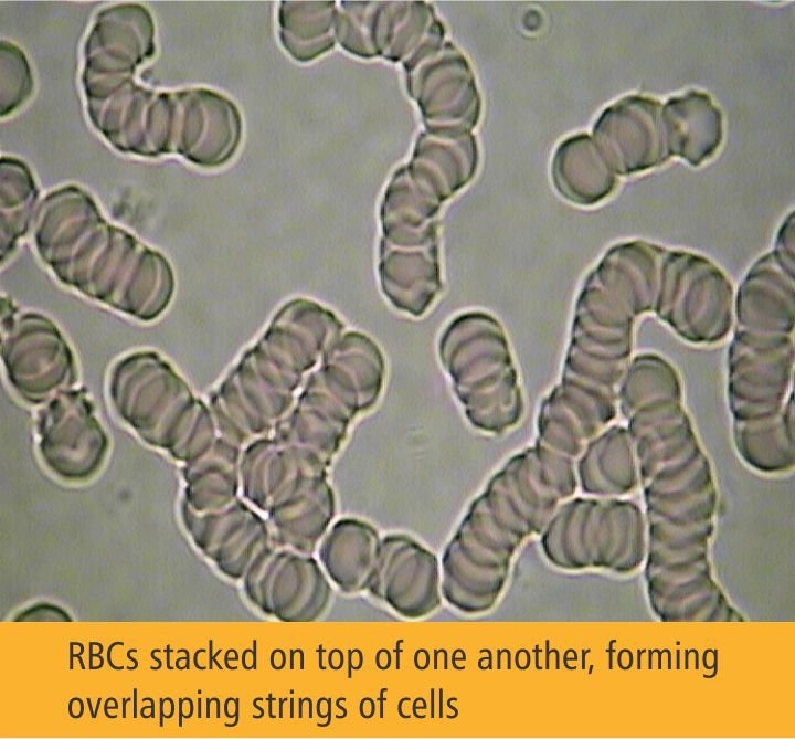
Red blood cells stacked like rolls of coins. Healthy erythrocytes repel each other and flow freely. When plasma proteins are elevated (often from inflammation, dehydration, or incomplete protein digestion), cells lose their negative surface charge and adhere. Marked rouleaux is one of the earliest signs of chronic low-grade inflammation, often visible long before CRP rises.
Ovalocytes (Elliptocytes)
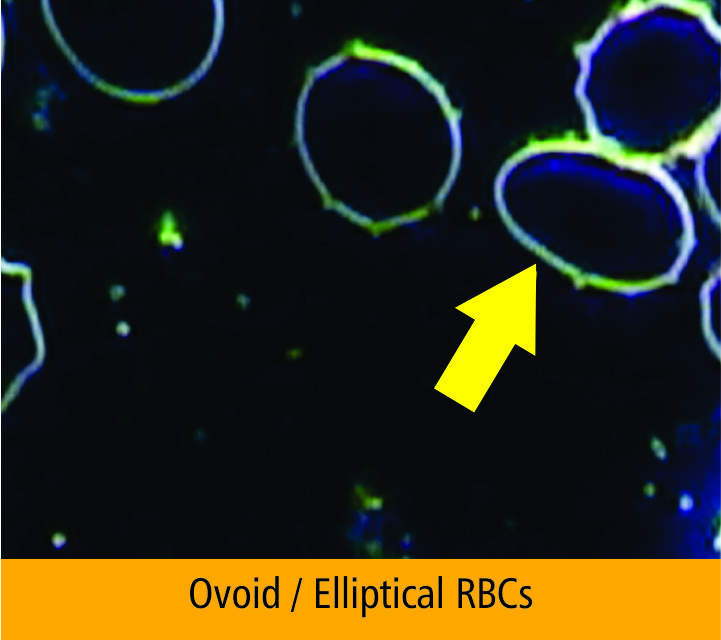
Egg or cigar-shaped red cells instead of round. A small percentage is normal. More than 25% of cells showing this morphology points toward B12, folate, or iron deficiency. Hereditary elliptocytosis is also possible, but in most adult reports the finding is nutritional and reversible with targeted supplementation.
Echinocytes
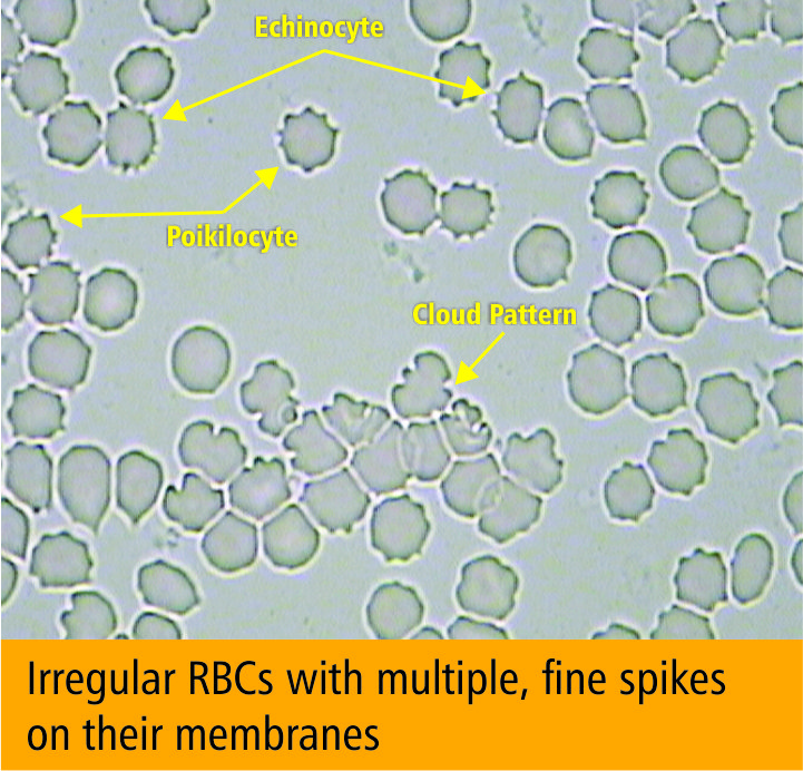
Red cells with short, evenly spaced spikes around the membrane (the «burr cell» appearance). Causes range from electrolyte imbalance and dehydration to uremia, liver disease, and oxidative stress. Transient echinocytes can appear after intense exercise. Persistent ones warrant follow-up on kidney and antioxidant status.
Target Cells
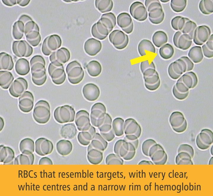
A bullseye pattern: a central dot of hemoglobin surrounded by a clear ring. Associated with thalassemia trait, iron deficiency anemia, liver disease, and post-splenectomy states. The cells have an abnormal surface-to-volume ratio, which changes how they navigate small vessels.
Acanthocytes
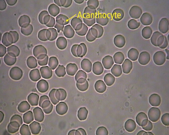
Irregular, thorn-like projections of varying length, distinct from the uniform spikes of echinocytes. Strong indicator of severe liver disease, lipid metabolism disorders (particularly abetalipoproteinemia), or post-splenectomy states. Even a few acanthocytes per field merit clinical attention.
Uric Acid Crystals
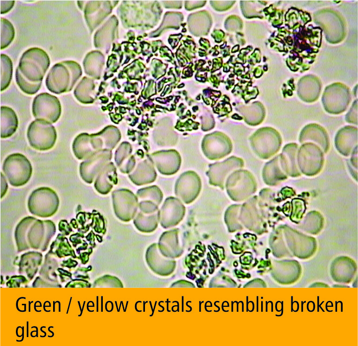
Sharp, needle-like or rhomboid crystals visible in the plasma, not the cells. Reflects systemic acid load and purine metabolism. Frequent appearance correlates with gout risk, kidney stress, and high-protein diets without adequate alkaline buffering.
Reading the report together. Every pattern in your Sangre Viva report comes with classification and clinical context delivered during the consultation. Patterns shift before standard labs flag them. That window is where prevention works.